Metaplasia, dysplasia, or carcinoma? #Esophagus #GIPath #PathTwitter #Pathology


#pathoshots #pathology #pathologyresidency #histopathology #microscopephotography #breastcancerawareness #breastpathology #usmle #neetpg #breastcancer #breast #BreastCancerAwarenessMonth instagram.com/reel/DAqLeKevr…
#pathoshots #pathology #pathologyresidency #pathologyresident #medicalstudent #medicaleducation #education #study #learning #skin #skinpathology #dermatopathology #histopathology instagram.com/reel/DASyylBvL…
Comment your Answers #pathoshots #pathology #pathologyresidency #pathologyresident #medicalstudent #medicaleducation #education #study #learning #thyroid #folliculartumor #follicularcancer #cancer #thyroidcancer #notcancer #Ft-UMP instagram.com/p/DAX-r8SPM2P/…
#pathoshots #pathology #pathologyresidency #pathologyresident #medicalstudent #medicaleducation #education #study #learning #histopathology #breastcancerawareness #breastpathology #breastcancer #breast #cancer #notcancer instagram.com/reel/DAd6po0v8…
🦠Xanthogranulomatous Pyelonephritis ⚠️ Mimic of Renal Cancer 📚 Kidney mass➡️Kidney🪨(often staghorn) ➡️ Obstructive uropathy➡️infection+inflammation ➡️fibrosis 🔬 Xanthoma, Inflammatory, Giant cells; Cholesterol clefts; Fibrosis 🩺 💊, Drainage, +/- Partial/Total Nephrectomy


🦴Aneurysmal Bone Cyst 📖Benign, multiloculated bone neoplasm w🩸filled cyst spaces 🩻Lytic lesion w/ well-defined margins + fluid-fluid levels 🗺️Often metaphysis of long🦴 📉Dx often first two decades 🧬USP6 (70%) 🔬Fibrous septa: bland fibroblasts + giant cells; mites common


Seminal vesicle nuclei: Too pleomorphic to be prostate cancer
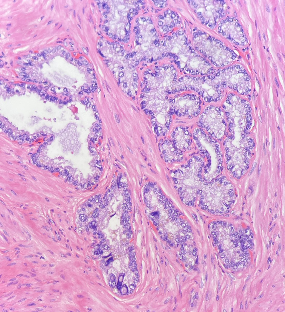
Testicular embryonal carcinoma with intratubular growth next to germ cell neoplasia in situ (GCNIS). #GUPath

Reactive mesothelial cells are so dramatic but so pretty 🥹. #cytologyisbeautiful #pathart #cytologyart #pathx #pathtwitter
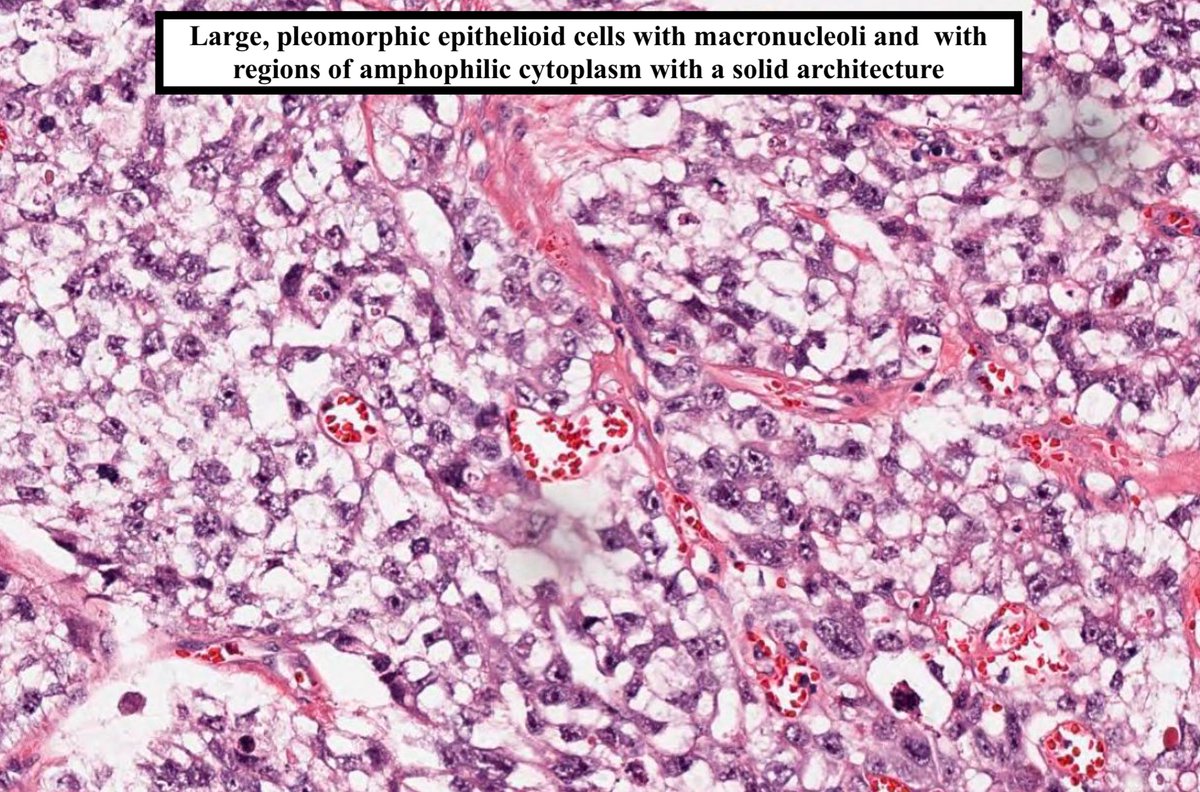
Mixed germ cell tumor with prominent intratubular embryonal carcinoma- often with comedonecrosis & Ca2+ (left part of image) #testis #Cancer #gupath #urology @UMichPath

Florid Intratubular embryonal carcinoma in a nice wedge shape. The invasive tumour is just present on the right. Note necrosis and expansion of the tubules. All of the fun of intraductal prostate carcinoma but none of the politics….


Intratubular embryonal carcinoma cuddling up against GCNIS. Note the tubule size difference, necrosis and cytology!

Intratubular Embryonal Carcinoma in a Mixed Germ Cell Tumor https://t.co/QwRPlh9eRb


Findings for embryonal carcinoma in cytologic preparations include tight clusters and scattered individual tumor cells with pleomorphic nuclei with irregular nuclear contours and prominent nucleoli. Unlike seminoma, a tigroid background is not seen. #GUpath

Embryonal Carcinoma • #2 most prevalent testicular germ cell tumor (after seminoma) • Peak incidence is 30 y/o (10 years younger than seminoma) • Most common element seen in mixed germ cell tumor. • IHC: CD30 and AE1/AE3 + Image Credit: Kelly Hall, MD #pathagonia

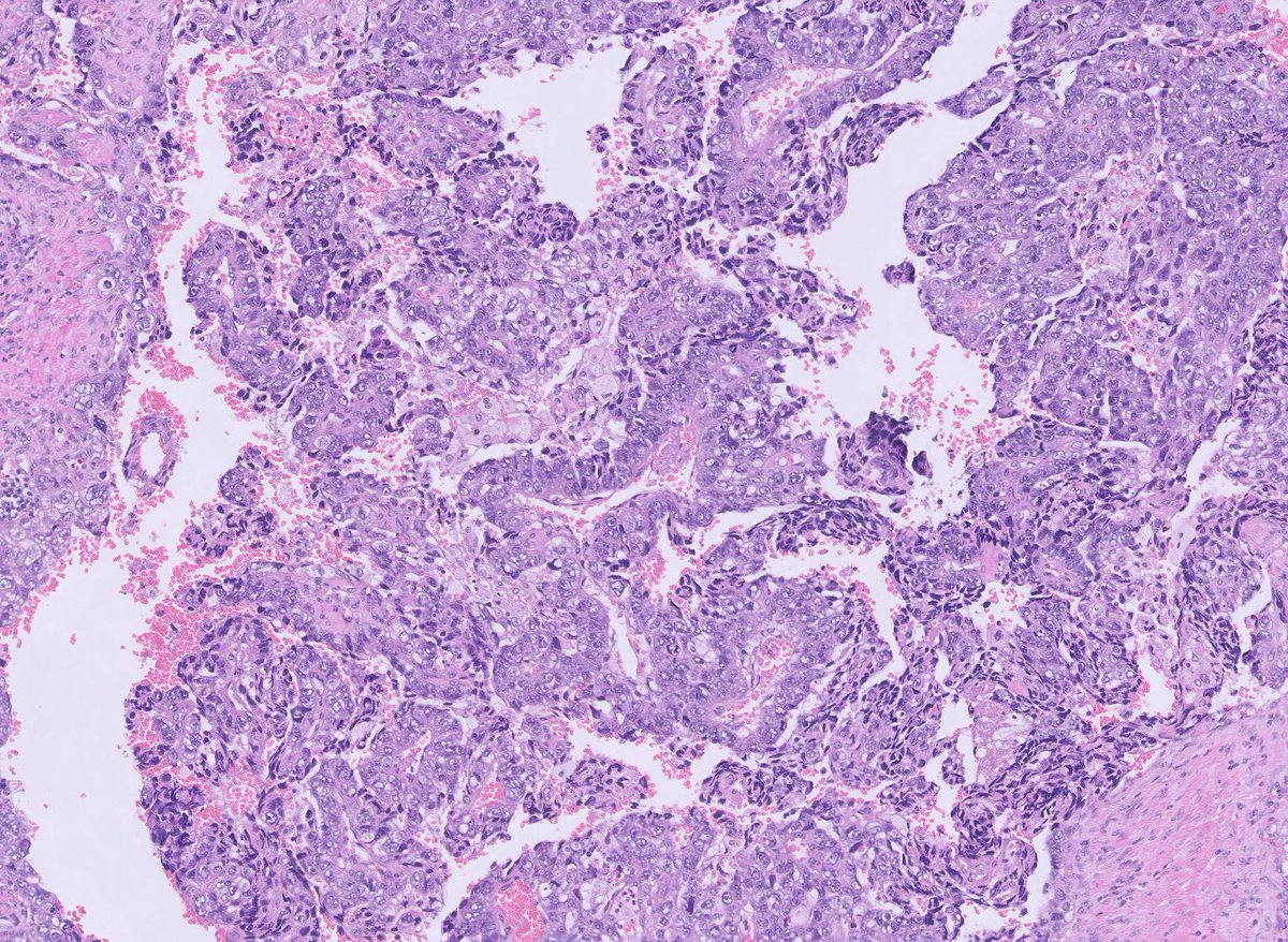
Florid Intratubular Embryonal carcinoma. ( and a bit of invasive EC!)


37 y/o M with hx of embryonal carcinoma of testis, now lymph node masses. See WSI, order stains, and check dx here: drdoubleb.com/crowdunknowns/…
United States Trends
- 1. #SmackDown 79,7 B posts
- 2. CM Punk 19,1 B posts
- 3. Paul Heyman 8.095 posts
- 4. Khalid 26,6 B posts
- 5. Jared McCain 12,9 B posts
- 6. Chaz Lanier 1.677 posts
- 7. Creighton 4.528 posts
- 8. #OPLive 2.626 posts
- 9. Kendrick 823 B posts
- 10. Bayley 4.757 posts
- 11. Bianca 17,5 B posts
- 12. #BlueBloods 2.147 posts
- 13. MSNBC 281 B posts
- 14. Wiseman 3.959 posts
- 15. #AEWRampage 2.750 posts
- 16. #loveafterlockup 1.892 posts
- 17. OG Bloodline 9.726 posts
- 18. Gorka 16,5 B posts
- 19. Kevin Owens 3.722 posts
- 20. War Games 13,6 B posts
Something went wrong.
Something went wrong.


