MSS India
@MSKSocietyIndiaThe Musculoskeletal Society, India is a nonprofit association of Musculoskeletal Radiologists dedicated to teaching & spreading awareness about MSK Radiology
Peripheral nerve imaging Radial nerve - Posterior cord of the brachial plexus (C5-C8, T1) Identify - spiral groove (easiest and consistent) Follow - up and down up to the bifurcation into superficial radial nerve and posterior interosseous nerve Diagnosis - spiral groove…

Peripheral nerve imaging Superficial branch of the radial nerve - pure sensory nerve Cheiralgia paresthetica, also known as Wartenberg’s syndrome or superficial radial nerve entrapment, is a condition that involves compression or entrapment of the superficial branch of the…

Consent ✅ Trail runner with lateral ankle pain What is the problem & the 'sign'?

The Joint Effort: MSK Quiz. 🥳 This segment is designed to challenge and enhance your diagnostic skills, offering a unique platform to test your knowledge in musculoskeletal radiology . We invite radiologists, clinicians, and medical professionals to contribute questions and…


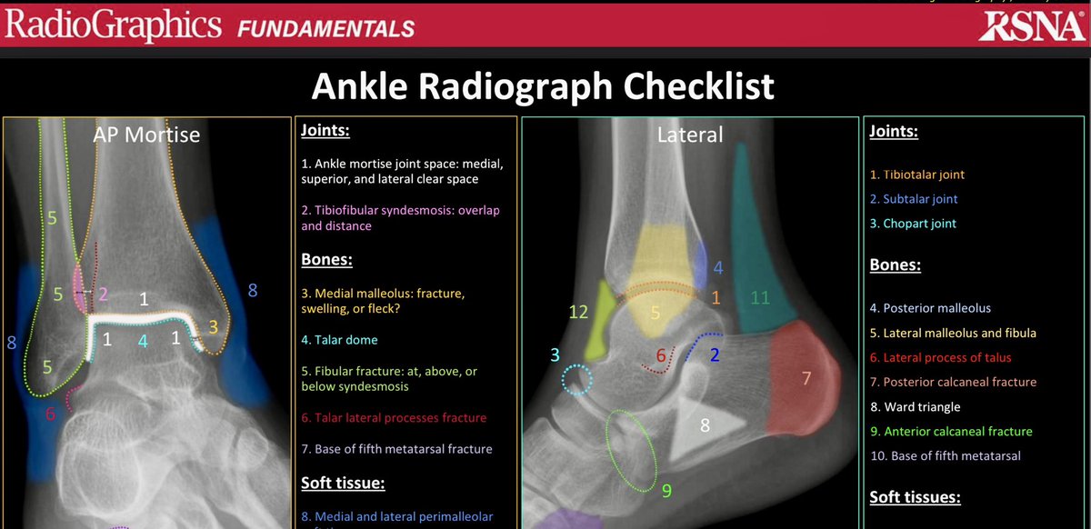
Don’t forget the base of the 5th Metatsrsal on the lateral ankle radiograph. Ankle radiograph checklist —on-call tips


Peripheral nerve imaging Posterior interosseous nerve - pure motor nerve Identify - between superficial and deep fibers of supinator muscle PIN entrapment - arcade of Frohse (between superficial and deep fibers of supinator), may have prominent adjacent vessels leading to…

Acute odynophagia in the ER. Don't forget Longus Colli calcicif tendinosis. Two patients on yesterday’s on-call… —on-call tips


Acute shoulder pain in the ER. Subcortical enthesopathic cystic changes of the greater tuberosity. Calcific tendinosis of the rotator cuff with an extension of calcification to the subcortical cystic lesions possibly suggests osseous involvement. -On-call cases


🟠 Dynamic ultrasound assessment for insertional achilles tendinopathy: the COcco-RIcci (CORI) sign Access and learn more 👉 rdcu.be/dYZ4y #SkeletalRadiology #MSKrad #radres #radiology #ultrasound

Another case of Trapped periosteum..similar to one we saw earlier in ournpediatric series week!
Trapped Periosteum: Diagnosis A complication of physeal fractures is the entrapment of the periosteum at the level of the fracture line in the physis, generally produced by a hyperextension mechanism with valgus or varus stress. The great majority are related to Salter-Harris…
⭐️ MR neurography is in another level with amazing images from very small nerves like in this Medial Antebrachial Cutaneous Nerve transection with a stump neuroma. @elihag @Aspetar #mrneurography #peripheralnerves #mskrad #radresidents


High-resolution ultrasound in the evaluation of the adult hip pmc.ncbi.nlm.nih.gov/articles/PMC10…


Skeletal Radiology November issue: 🟢 Review Article | rdcu.be/dYZON 🔴 Scientific Article | rdcu.be/dYZPk #SkeletalRadiology #MSKrad #radres #radiology #orthopedics

Peripheral nerve imaging Quick pro tips for seeing the unseen - peripheral nerves of the upper extremity One nerve a day! For today - Knobology and probe selection! #nerveimaging #mskrad #mskradiology #mskusg #loveformsk #nerves #injuries #msk #mskRad #FOAMRad #FOAMMed…

Hill-Sachs defects are best seen on AP shoulder radiographs in internal rotation. Anterior shoulder dislocation pre-reduction. —on-call cases


Subchondral lucency of the talar dome seen in AP view after talar neck # is a favorable sign (Hawkins sign). This indicates that there is sufficient vascularity in the talus, and is therefore unlikely to develop avascular necrosis of the talar dome later...

I'm excited to share that my third first-author article in @RadioGraphics has been published! The RadioGraphics journal has played a crucial role in my development as an academic radiologist and educator in radiology. Thank you to @drdevrad for your enthusiasm, commitment,…

🚨 In a few hours! Join us today at 12:00 pm CST for an insightful ACORE Webinar on "High-frequency Ultrasound of Common Wrist and Hand Pathologies." Dr. Ramy Mansour, MSK Consultant Radiologist from the University of Ottawa, will be presenting, with Dr. Raj Chari from Oxford…

Saga of pitfalls around the wrist... #knee #postop #xray #msk #mskRad #FOAMRad #FOAMMed #radiology #RadRes #medicine #orthotwitter #MSKImaging #Radiology #Education #Musculoskeletal #FoamRad #medtwitter @samrad77 @SkeletalRadiol @The_ASSR @RadioGraphics @ClinRadiology…

One view is NO view. Crescent sign: Subchondral insufficiency fracture.


United States Trends
- 1. #PaulTyson 102 B posts
- 2. Goyat 11,9 B posts
- 3. #SmackDown 11,8 B posts
- 4. Rosie Perez 1.488 posts
- 5. Cedric 5.528 posts
- 6. Bayley 5.074 posts
- 7. #netfilx N/A
- 8. #NetflixBoxing N/A
- 9. Cam Thomas 2.207 posts
- 10. B-Fab 4.037 posts
- 11. CJ Cox N/A
- 12. Max Christie 1.212 posts
- 13. LA Knight 2.752 posts
- 14. Purdue 4.923 posts
- 15. #FightNight 4.038 posts
- 16. Hukporti 1.085 posts
- 17. Karoline 39,1 B posts
- 18. Kevin Love 1.917 posts
- 19. Michin 3.407 posts
- 20. End 1Q N/A
Something went wrong.
Something went wrong.


