Rajesh S
@Dr_RajeshSDiagnostic Neuroradiologist #firstgenerationdoctor | Most/All the tweets are radiology related| Instagram @buddingradiologist #FOAMrad #Medtwitter
Similar User

@draceciferrario

@drvenkimdrd

@CasesCookyJar

@msk_munoz

@TheENRS

@Radquarters

@samrad77

@drharunyildiz

@tabby_kennedy

@drSurjthVattoth

@ManavendraUpad1

@learnneurorad

@CoolAsANeuroRad

@CafeRoentgen

@unc_neurorads
🧠Petrous apex cholesterol granuloma🧀 Middle aged with h/o COM T1 hyperintense(due to cholesterol component&methemoglobin)lesion centered in the petrous apex,T2/FLAIR central high signal with low hemosiderin rim Other skull base tumours&thrombosed aneurysm can mimic #neurorad

Visiting Goa for the first time to attend this wonderful Paediatric Neuroradiology conference. After completing part 1&2 virtually,excited to meet all the renowned teachers in person for the final part SPIN 2024.Beautiful line up of lectures at the sea cost, Great treat🎉#SPIN24


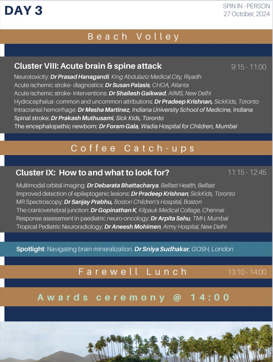
'Tis the Oktoberfest of Pediatric Neuroradiology in a month from now! In 30 days exactly we will say Namaste to our incredible faculty, and our equally incredible delegates. The Fellowship of #SPIN beckons....

🧠Diffuse hyperdensity of the intracranial vessels on NCCT looks exactly like post contrast images.Not to be mistaken for thrombus Can be seen in patients with: Elevated hematocrit Polycythemia Severe dehydration This patient had cyanotic cong cardiac ds-TOF with high hematocrit


Excellent summary 👏of all you need to know about the Chiari malformations, From imaging workup till the treatment Type 0 to 1.5 are developmental alterations Vs Type 2 to 5 true malformations @RadioGraphics @JamieClarkeRad #neurorad #radres #Foamrad


Features of Chiari I and related deformities are shortening of the clivus and supra-occiput, tonsillar herniation, and crowding of the foramen magnum. Chiari I can be diagnosed with cerebellar tonsillar herniation of 3-5mm if supporting features (like syrinx) are present. #RGphx

🧠Non neoplastic corpus callosal lesions: Multiple sclerosis - Involves callososeptal interface Marchiafa Bignami - Involves central layers with relative sparing of the dorsal&ventral extremes(Sandwich) Susac syndrome -Spherical/round lesions in the center of CC(Snowball/Icicles)

🧠Knowing Juxta Vs Subcortical location is very important particularly in diagnosing MS & leukodystrophies. It is misunderstood & loosely used term by many Juxta/leukocortical: Involve &abuts the Ufibres Subcortical: Few mm away from the Ufibres @Radiopaedia #neurorad #radres


What's the odd of fish bone hitting the vertebral artery 🤯
#STROKE Images: Throat discomfort and hemoptysis after accidental ingestion of a fish bone – with penetration of the left vertebral artery. 🐟 #AHAJournals ahajrnls.org/3XiI1IZ

Calcified left MCA embolism🧠 Not to be mistaken for calcified granuloma/neoplasm in acute setting Calcified cerebral emboli are rare cause of ischemic stroke.Majority are from the calcified valve/ruptured calcified plaques If multiple-Salted pretzel sign may be seen #neurorad

United States Trends
- 1. Dolphins 40,6 B posts
- 2. #GoPackGo 9.198 posts
- 3. Datsun 8.521 posts
- 4. McDaniel 7.354 posts
- 5. Josh Jacobs 7.774 posts
- 6. #WinterAhead 144 B posts
- 7. Jordan Love 8.266 posts
- 8. Tulane 6.248 posts
- 9. Green Bay 10,5 B posts
- 10. Tyreek 6.647 posts
- 11. #TPNE 1.095 posts
- 12. Jayden Reed 3.586 posts
- 13. Grier 1.843 posts
- 14. #MIAvsGB 9.277 posts
- 15. Jason Garrett 1.317 posts
- 16. The Party Never Ends 13,3 B posts
- 17. Jonnu 2.773 posts
- 18. Achane 3.289 posts
- 19. bibby 1.095 posts
- 20. Mostert N/A
Who to follow
-
 Dra. Cecilia Ferrario
@draceciferrario -
 Venkatesh M☯️
@drvenkimdrd -
 Cases From the Cooky Jar
@CasesCookyJar -
 JP Munoz • MSK
@msk_munoz -
 The Eastern Neuroradiological Society
@TheENRS -
 Daniel Kowal, MD, RMSK | Radquarters
@Radquarters -
 Sameer Raniga
@samrad77 -
 Dr Harun Yıldız
@drharunyildiz -
 Tabby A. Kennedy, MD
@tabby_kennedy -
 Surjith Vattoth
@drSurjthVattoth -
 Manavendra Upadhyaya,MD
@ManavendraUpad1 -
 Divya Gunda
@learnneurorad -
 Rajan Jain, MD
@CoolAsANeuroRad -
 Cafe Roentgen
@CafeRoentgen -
 UNC Neuroradiology
@unc_neurorads
Something went wrong.
Something went wrong.


